What tests should be carried out in hospital after a stroke?

After a stroke, it is crucial to carry out certain examinations in hospital to identify the causes, assess the severity and plan appropriate treatment. In the best case scenario, these examinations take place in a stroke unit. In this blog post, you will find out which examinations should be carried out in hospital after a stroke and why they are important.
Contents:
- Imaging procedures: CT and MRI
- Blood tests: Complete blood count and blood sugar
- Doppler and duplex sonography: blood vessel examination
- Angiography: visualization of the blood vessels
- Electrocardiogram (ECG): Checking the heart rhythm
- Neuropsychological tests: assessment of cognitive functions
Imaging procedures: CT and MRI
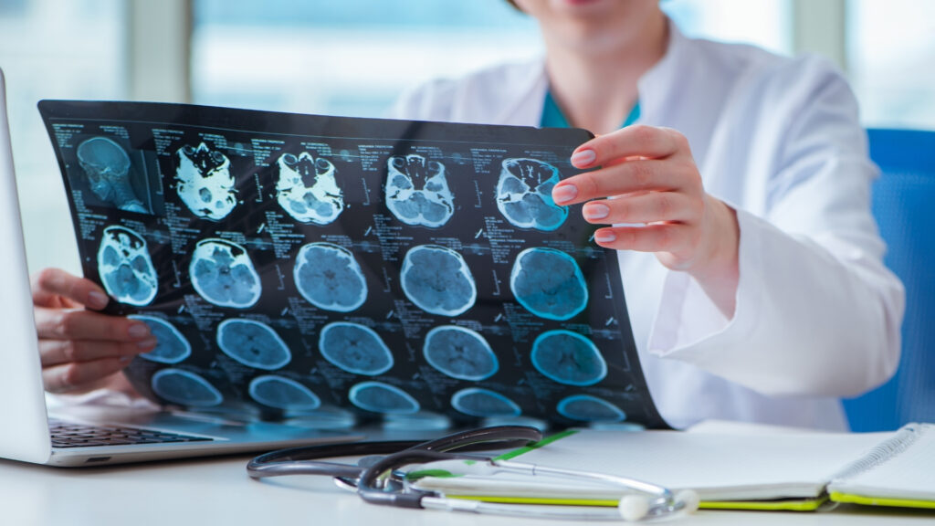
Imaging procedures such as computed tomography (CT) and magnetic resonance imaging (MRI) are essential to determine the extent of the stroke. CT scans provide quick results and can help to identify a brain hemorrhage or damage. MRI scans provide more detailed images and help to detect ischemic strokes and smaller lesions. Knowing the cause of the stroke or what type of stroke it is is crucial for treatment. You can read more about the processes in the brain and the different types of stroke here.
Both methods can display the brain as if it were cut into thin layers and thus precisely depict the various regions, which is very helpful for diagnosis. The procedures use different methods.
Computed tomography (CT) uses X-rays to create detailed cross-sectional images of the body. The treating physicians will inform you about the possible risks. During a CT scan, the patient is placed on a table that moves through a ring-shaped scanner.
Blood tests: Complete blood count and blood sugar
Blood tests are crucial in order to clarify possible causes and risk factors for a stroke. A complete blood count can be used to detect anemia (also anemia) or infections in the body. In anemia, the haemoglobin content, i.e. the number of red blood cells, is reduced. This makes it harder for oxygen to be transported through our body.
Blood sugar levels can also provide indications of diabetes. All three factors are risk factors for another stroke and should be treated. The blood is also tested for coagulation disorders. This is because strokes are often caused by blood clots (so-called thrombi). You can find out more about what happens in the brain during a stroke here.
Doppler and duplex sonography: blood vessel examination
Doppler sonography measures the blood flow in the arteries and veins. This helps to identify constrictions or blockages in the blood vessels that could have led to a stroke. It is particularly important to check for deposits and blood clots in the carotid arteries.
Doppler sonography uses ultrasound to visualize and measure the blood flow in the vessels. The doctor or technician applies a special gel to the patient’s skin to create good contact between the ultrasound probe and the skin. The ultrasound probe is then gently moved over the skin to observe the blood flow in the blood vessels.
Duplex sonography combines Doppler sonography with conventional ultrasound images to assess both the blood flow and the structure of the vessels. Similar to Doppler sonography, a gel is applied to the skin and the ultrasound probe is used to create images of the blood vessels. This enables a more detailed assessment of constrictions, deposits or other anomalies in the blood vessels. For example, deposits in the vessels can also be made visible.
Angiography: visualization of the blood vessels
Angiography is a radiological examination that enables high-resolution imaging of the blood vessels. It can be used to create detailed images of the brain vessels and identify constrictions, aneurysms, blood clots or other vascular abnormalities. This helps those treating the patient to determine the exact cause of the stroke and to plan the appropriate treatment.
By combining these different imaging techniques, doctors can make a comprehensive assessment of the blood vessels in the brain and identify potential causes of the stroke. This enables a precise diagnosis to be made and an individual treatment plan to be developed for the patient.
Electrocardiogram (ECG): Checking the heart rhythm
An electrocardiogram (ECG) is performed to check the heart rhythm. Certain cardiac arrhythmias such as atrial fibrillation can increase the risk of blood clots and lead to a stroke.
The patient usually has to expose the upper body so that the electrodes can be attached to the chest. The electrodes are connected to an ECG device that records the electrical signals of the heart. The examination is painless and the duration of the ECGs is determined by the practitioners. During the examination, the patient should lie as still as possible, as movements can influence the results. The ECG shows the heart rate, the heart rhythm and can detect abnormalities such as arrhythmias or signs of a heart attack.
Neuropsychological tests: assessment of cognitive functions
The early identification of causes and risk factors enables doctors to take targeted measures to prevent future strokes.
Actively ask for these examinations and insist that they are all carried out. Comprehensive clarification of all risk factors and sequelae is crucial for rehabilitation and prevention.
As a rule, patients do not need to make any special preparations for these examinations. However, it is important to provide the medical staff with all relevant information about existing illnesses, medication or allergies in order to ensure the safety and accuracy of the examinations.

